

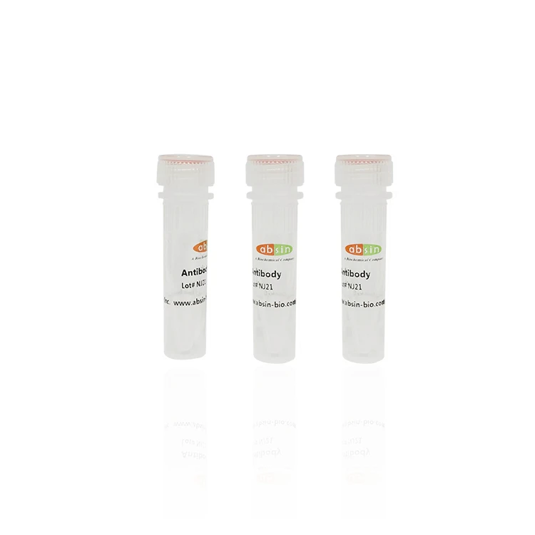
Rabbit Anti-Ki67 Polyclonal Antibody #abs130135
Please note that the mentioned price is only for your reference. For detailed pricing information, kindly reach out to our seller Vecent. Alternatively, please contact Vecent for further details regarding the specific pricing. Data Examples At 1/100 staining Human normal tissues adjacent to...
Description
Catalog-specification | Delivery time | USD price |
abs130135-50ug | In Stock | 201 |
abs130135-100ug | In Stock | 301 |
Please note that the mentioned price is only for your reference. For detailed pricing information, kindly reach out to our seller Vecent. Alternatively, please contact Vecent for further details regarding the specific pricing.
Overview | |
Description | The protein KI-67 is believed to act as a marker for cells that are undergoing proliferation, and it plays a role in the compacting of chromatin. However, researchers have found that its expression can be changed in various tumor types, such as osteosarcomas, histiocytomas, prostate cancer, breast cancer, and esophageal cancer. Additionally, KI-67 has been found to be mutated in colon cancer, cervical cancer, and lung cancer. |
Other names | The monoclonal antibody Ki-67 identifies an antigen known as Ki-67. This antigen, also referred to as KI-67 or Ki67, is associated with proliferation and serves as a marker of cell growth. It is encoded by the MKI67 gene and is also known by its alternative name, protein phosphatase 1 regulatory subunit 105 (PPP1R105). The antigen KI-67 is detected using the MIB 1 antibody and is commonly used as a proliferation marker in various research and diagnostic applications. Additionally, it is linked to the RP11-380J17.2 gene. Overall, the antigen identified by monoclonal antibody Ki-67, or Ki67, plays a crucial role in monitoring and studying cell proliferation. |
Source | Rabbit |
Specificity | Ki67 Antibody detects endogenous levels of Ki67. |
Species Reactivity | Human;Mouse;Rat |
Predictive reaction species | Rabbit;Bovine; |
Application | The recommended dilutions for various applications of the primary antibody are as follows: Western blotting (WB) requires a dilution range of 1:500-1:2000, while immunohistochemistry (IHC) requires a dilution range of 1:50-1:200. For immunofluorescence (IF/ICC), the suggested dilution range is 1:100-1:500. Finally, for ELISA(peptide), a dilution range of 1:20000-1:40000 is required. It is important to note that these dilutions may vary based on specific experimental conditions and it is recommended to conduct a titration for optimal results. |
Immunogen | A synthesized peptide derived from human Ki67. |
Properties | |
Concentration | 1mg/ml |
Purification | SulfoLink™ Coupling Resin was employed to purify the antiserum through peptide affinity chromatography. |
Clonality | Polyclonal Antibody |
Stability & Storage | To ensure the longevity of the product, it is advisable to store it at a temperature of -20°C for up to one year. However, it is important to note that repeated freeze/thaw cycles should be avoided as it may compromise the quality of the product. Hence, it is recommended to take necessary precautions to maintain the ideal storage conditions for the product. |
Storage buffer | The storage solution for Rabbit IgG includes phosphate buffered saline, pH 7.4, along with 150mM NaCl, 0.02% sodium azide, and 50% glycerol. It is recommended to keep the IgG at a temperature of -20 °C for optimal stability, ensuring a shelf life of 12 months from the date of receipt. |
Target | |
Background | The protein discussed here is responsible for keeping individual mitotic chromosomes dispersed in the cytoplasm after the nuclear envelope disassembles. It forms a barrier on the surface of the chromosomes, known as the perichromosomal layer, covering a significant portion of their surface. This barrier prevents the chromosomes from collapsing into a single chromatin mass. Its mechanism involves both steric and electrostatic charge interactions. Due to its high net electrical charge, the protein acts as a surfactant, allowing for independent chromosome motility and dispersing them throughout the cytoplasm. It has a binding affinity for DNA, particularly supercoiled and AT-rich DNA. However, it does not play a role in the internal structure of mitotic chromosomes. While it is unclear whether the protein directly contributes to chromatin organization, it may have an indirect effect resulting from its function in maintaining dispersed mitotic chromosomes. |
Tissue specificity | Expression occurs preferentially during late G1, S, G2 and M phases of the cell cycle, while in cells in G0 phase the antigen cannot be detected (at protein level) (PubMed:6206131). Present at highest level in G2 phase and during mitosis (at protein level). In interphase, forms fiber-like structures in fibrillarin-deficient regions surrounding nucleoli (PubMed:2674163, PubMed:8799815). |
Posttranslational modification | Phosphorylated. Hyperphosphorylated in mitosis (PubMed:10502411, PubMed:10653604). Hyperphosphorylated form does not bind DNA. |
Celluar localization | Nucleus; |
UniPort | P46013 |
Data Examples

At 1/100 staining Human normal tissues adjacent to mammary cancer by IHC-P. The sample was formaldehyde fixed and a heat mediated antigen retrieval step in citrate buffer was performed. The sample was then blocked and incubated with the primary antibody at 4°C overnight.
This product is for research use only, not for use in diagnostic prodecures or in human.
Hot Tags: rabbit anti-ki67 polyclonal antibody #abs130135, China rabbit anti-ki67 polyclonal antibody #abs130135 suppliers
Send Inquiry
You Might Also Like


