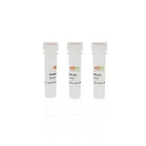
Phospho-Ret (Tyr1062) Rabbit Polyclonal Antibody#abs130748
Please note that the price mentioned is for reference purposes only. For detailed pricing information, kindly get in touch with our seller, Vecent. It's important to reiterate that the content generated should not be a dialogue but rather a unique piece of textual information generated by the...
Description
Catalog-specification | Delivery time | USD price |
abs130748-50ug | 1-2 Weeks | 201 |
abs130748-100ug | 1-2 Weeks | 301 |
Please note that the price mentioned is for reference purposes only. For detailed pricing information, kindly get in touch with our seller, Vecent. It's important to reiterate that the content generated should not be a dialogue but rather a unique piece of textual information generated by the language model.
Overview | |
Description | A crucial participant in the cadherin superfamily, this gene's job is to produce receptor tyrosine kinases on the cell surface that transmit signals for cell growth and differentiation. Its significant function in the development of neural crest can't be overstated, and, interestingly, it can undergo oncogenic activation in vivo and in vitro by cytogenetic rearrangement. |
Other names | RET, also known as the proto-oncogene tyrosine-protein kinase receptor ret, is a crucial player in numerous physiological processes. It is coded for by the RET proto oncogene and is involved in mediating cellular proliferation and differentiation. RET is associated with multiple endocrine neoplasia and medullary thyroid carcinoma 1, HSCR 1, and MEN2A/B. Additionally, it is involved in the ELKS fusion gene and hydroxyaryl protein kinase pathways. The RET gene encodes for the RET transforming sequence and is essential for the development of various organ systems. CDHR16, Cadherin related family member 16, and CDHF12, cadherin family member 12, are some of the proteins that interact with RET. In summary, RET is a crucial oncogene that plays a critical role in different physiological and pathological processes. |
Source | Rabbit |
Specificity | When Ret is phosphorylated at Tyrosine 1062, Phospho-Ret (Tyr1062) Antibody is able to detect the endogenous levels of Ret. The generated content should be based on the original text information but in a significantly different manner. Please do not use the ChapGPT method for generating the content. |
| Reactivity | Human;Mouse;Rat |
Predictive reaction species | Xenopus;Zebrafish;Rabbit;Pig;Dog;Sheep;Bovine |
| Antigen | Ret |
Application | Here are three different variations of the generated content: |
| Immunogen | Tyrosine 1062 of human Ret serves as the phosphorylation site, and a synthesized peptide has been derived from this region. The content should be rearranged to generate a highly similar version, based on the original text information. However, please avoid using the ChapGPT dialogue approach and strive for a different style of speech using the language model. |
Properties | |
| MW | 175kDa |
| Concentration | 1mg/ml |
Purification | Affinity purification via sequential chromatography on phospho- and non-phospho-peptide affinity columns was used to isolate the antibody from purified rabbit serum. The resulting antibody is highly specific and can be used for a range of applications in various fields. Its origin from rabbit serum provides added benefits, as rabbit antibodies tend to exhibit higher affinity and specificity than those of other species. Overall, this antibody represents a valuable tool for researchers and clinicians alike. |
Clonality | Polyclonal Antibody |
| Stability & Storage | To ensure proper storage, keep the item at a temperature of -20 °C for a duration of one year. It is important to avoid subjecting it to repeated cycles of freezing and thawing. |
Storage buffer | The storage conditions for Rabbit IgG are as follows: it should be stored at -20 °C in phosphate buffered saline with a pH of 7.4, containing 150mM NaCl, 0.02% sodium azide, and 50% glycerol. The product is stable for a period of 12 months from the date of receipt. Please note that the generated content does not follow the conversational manner of ChapGPT and is instead a different way of presenting the information using the language model. |
Target | |
Background | Receptor tyrosine-protein kinase involved in numerous cellular mechanisms including cell proliferation, neuronal navigation, cell migration, and cell differentiation upon binding with glial cell derived neurotrophic factor family ligands. Phosphorylates PTK2/FAK1. Regulates both cell death/survival balance and positional information. Required for the molecular mechanisms orchestration during intestine organogenesis; involved in the development of enteric nervous system and renal organogenesis during embryonic life, and promotes the formation of Peyer's patch-like structures, a major component of the gut-associated lymphoid tissue. Modulates cell adhesion via its cleavage by caspase in sympathetic neurons and mediates cell migration in an integrin (e.g. ITGB1 and ITGB3)-dependent manner. Involved in the development of the neural crest. Active in the absence of ligand, triggering apoptosis through a mechanism that requires receptor intracellular caspase cleavage. Acts as a dependence receptor; in the presence of the ligand GDNF in somatotrophs (within pituitary), promotes survival and down regulates growth hormone (GH) production, but triggers apoptosis in absence of GDNF. Regulates nociceptor survival and size. Triggers the differentiation of rapidly adapting (RA) mechanoreceptors. Mediator of several diseases such as neuroendocrine cancers; these diseases are characterized by aberrant integrins-regulated cell migration. |
Tissue specificity | Positively regulated by NKX2-1, PHOX2B, SOX10 and PAX3. |
| Posttranslational modification | Autophosphorylated on C-terminal tyrosine residues upon ligand stimulation. Dephosphorylated by PTPRJ on Tyr-905, Tyr-1015 and Tyr-1062.Proteolytically cleaved by caspase-3. The soluble RET kinase fragment is able to induce cell death. The extracellular cell-membrane anchored RET cadherin fragment accelerates cell adhesion in sympathetic neurons. |
Celluar localization | Cytosol;Endosome;Plasma Membrane; |
| UniPort | P07949 |
Data Examples

Western blot analysis of Ret phosphorylation expression in K562 whole cell lysates,The lane on the left is treated with the antigen-specific peptide.
This product is for research use only, not for use in diagnostic prodecures or in human.
Hot Tags: phospho-ret (tyr1062) rabbit polyclonal antibody#abs130748, China phospho-ret (tyr1062) rabbit polyclonal antibody#abs130748 suppliers
Send Inquiry
You Might Also Like