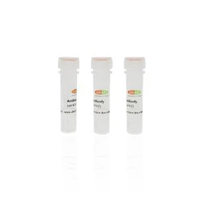
Rabbit Anti-Cofilin Polyclonal Antibody #abs130555
Please note that the price provided above is for your reference only. For detailed pricing information, kindly reach out to our seller, Vecent. We encourage you to contact Vecent to obtain accurate and up-to-date pricing details. Data Examples Western blot analysis on Jurkat cell lysate using...
Description
Catalog-specification | Delivery time | USD price |
abs130555-50ug | 1-2 Weeks | 201 |
abs130555-100ug | 1-2 Weeks | 301 |
Please note that the price provided above is for your reference only. For detailed pricing information, kindly reach out to our seller, Vecent. We encourage you to contact Vecent to obtain accurate and up-to-date pricing details.
Overview | |
Description | Cofilin 1 is a protein found in the cytoskeleton that plays a crucial role in regulating actin depolymerization. It exhibits a significantly higher affinity, about 5-10 times more, for ATP-actin monomers compared to cofilin-1. Interestingly, it has been suggested that cofilin-1 may actually promote the assembly of actin filaments rather than their disassembly. There are two different variations of cofilin-1 resulting from alternative splicing events. |
Other names | One of the proteins that plays a crucial role in the functioning of our muscles is cofilin. In humans, there are two different types of cofilin - cofilin 1 and cofilin 2. Cofilin 1 is primarily found in non-muscle tissues, while cofilin 2 is specifically located in muscle tissue. |
Source | Rabbit |
Specificity | The detection of endogenous levels of Cofilin can be achieved with the use of Cofilin Antibody. In order to generate similar content, the arrangement of the words and phrases can be altered without changing the original information. |
Species Reactivity | Human;Mouse;Rat |
Predictive reaction species | Zebrafish;Chicken;Rabbit;Xenopus;Dog;Horse;Bovine;Sheep;Pig |
Application | The suggested dilution range for using Western Blotting is 1:500-1:2000. The dilution range for Immunofluorescence and Immunocytochemistry is 1:100-1:500. For ELISA with peptide, the recommended dilution is 1:20000-1:40000. Please note that these dilution ranges are based on the original text information and should be followed accordingly. |
Immunogen | A synthesized peptide derived from human Cofilin. |
Properties | |
Concentration | 1mg/ml |
Purification | SulfoLink™ Coupling Resin was employed to purify the antiserum through peptide affinity chromatography. |
Clonality | Polyclonal Antibody |
Stability & Storage | To maintain the quality of the product, it is recommended to store it at a temperature of -20 °C for up to a year. It is important to avoid exposing it to repeated freeze and thaw cycles, as this may compromise its efficacy and stability. Keep in mind that proper storage is crucial for the preservation of the product's integrity and functionality. |
Storage buffer | The storage requirements for Rabbit IgG are as follows: It should be stored at -20 °C in a solution containing phosphate buffered saline with a pH of 7.4, 150mM NaCl, 0.02% sodium azide, and 50% glycerol. This solution ensures the stability of Rabbit IgG for a period of 12 months from the date of receipt. |
Target | |
Background | This protein is involved in the reversible regulation of actin polymerization and depolymerization in response to changes in pH. Its ability to depolymerize F-actin is controlled by its association with CSPR3, as reported in a study (PubMed:19752190). The protein can bind to both G-actin and F-actin, with a 1:1 ratio of cofilin to actin. It is a key component of actin rods found within the nucleus and cytoplasm. Its presence is essential for maintaining muscle integrity, and it may also play a role in the alteration of alpha-actin forms during the early stages of sarcomere remodeling in the postnatal period (By similarity). |
Tissue specificity | Isoform CFL2b is expressed predominantly in skeletal muscle and heart. Isoform CFL2a is expressed in various tissues. |
Posttranslational modification | The phosphorylation of Ser-24 may prevent recognition of the nuclear localization signal. |
Celluar localization | Cytoskeleton;Extracellular region or secreted;Nucleus; |
UniPort | Q9Y281/P23528 |
Data Examples

Western blot analysis on Jurkat cell lysate using Cofilin Antibody
This product is for research use only, not for use in diagnostic prodecures or in human.
Hot Tags: rabbit anti-cofilin polyclonal antibody #abs130555, China rabbit anti-cofilin polyclonal antibody #abs130555 suppliers
Send Inquiry
You Might Also Like